The Sacrum Bone
Explore the anatomy, structure, and role of the sacrum bone with Innerbody's interactive 3D model.

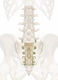
The sacrum is a large wedge shaped vertebra at the inferior end of the spine. It forms the solid base of the spinal column where it intersects with the hip bones to form the pelvis. The sacrum is a very strong bone that supports the weight of the upper body as it is spread across the pelvis and into the legs. Developmentally, the sacrum forms from five individual vertebrae that start to join during late adolescence and early adulthood to form a single bone by around the age of thirty. A ridge of tubercles along the posterior surface of the sacrum represents the spinous processes of these fused bones.
At its wide superior end, the sacrum forms the fibrocartilaginous lumbosacral joint with the fifth lumbar vertebra above it. The sacrum tapers to a point at its inferior end, where it forms the fibrocartilaginous sacrococcygeal joint with the tiny coccyx (tail bone). On the left and right lateral sides the sacrum forms the sacroiliac joints with the ilium of the hip bones to form the rigid pelvis. Many ligaments bind the sacroiliac joints together tightly to reduce motion and solidify the pelvis. Along its anterior surface the sacrum is concave to provide a larger space within the pelvic cavity. The female sacrum is shorter, wider, and curved more posteriorly than the male sacrum to provide more room for the passage of the fetus through the birth canal during childbirth.
Many nerves of the cauda equina at the inferior end of the spinal cord pass through the sacrum. These nerves enter the sacrum from the vertebral foramen of the lumbar vertebrae through the tunnel-like sacral canal. From the sacral canal these nerves branch out and exit the sacrum through four pairs of holes on the sides of the canal called the sacral foramina or through the sacral hiatus at the inferior end of the canal.
The sacrum serves several important functions in the skeletal, muscular, nervous, and female reproductive systems. Acting as the keystone of the pelvis, the sacrum locks the hip bones together on the posterior side and supports the base of the spinal column as it intersects with the pelvis. Several key muscles of the hip joint, including the gluteus maximus, iliacus and piriformis, have their origins on the surface of the sacrum and pull on the sacrum to move the leg. The sacrum also surrounds and protects the spinal nerves of the lower back as they wind their way inferiorly toward the end of the trunk and into the legs. Finally, the sacrum helps to form the pelvic cavity that supports and protects the delicate organs of the abdominopelvic cavity and provides space for a fetus to pass through during childbirth.


