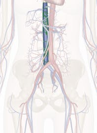
The Inferior Vena Cava
Explore Innerbody's 3D anatomical model of the inferior vena cava, the largest vein in the human body.

The inferior vena cava is the largest vein in the human body. It collects blood from veins serving the tissues inferior to the heart and returns this blood to the right atrium of the heart. Although the vena cava is very large in diameter, its walls are incredibly thin due to the low pressure exerted by venous blood.
The inferior vena cava forms at the superior end of the pelvic cavity when the common iliac veins unite to form a larger vein. From the pelvis, the inferior vena cava ascends through the posterior abdominal body wall just to the right of the vertebral column. Along its way through the abdomen, blood from the internal organs joins the inferior vena cava through a series of large veins, including the gonadal, renal, suprarenal and inferior phrenic veins. The hepatic vein provides blood from the digestive organs of the abdomen after it has passed through the hepatic portal system in the liver. Blood from the tissues of the lower back, including the spinal cord and muscles of the back, enters the vena cava through the lumbar veins. Many smaller veins also provide blood to the vena cava from the tissues of the abdominal body wall. Upon reaching the heart, the inferior vena cava connects to the right atrium on its posterior side, inferior to the connection of the superior vena cava.
The inferior vena cava and its tributaries drain blood from the feet, legs, thighs, pelvis and abdomen and deliver this blood to the heart. Many one-way venous valves help to move blood through the veins of the lower extremities against the pull of gravity. Blood passing through the veins is under very little pressure and so must be pumped toward the heart by the contraction of skeletal muscles in the legs and by pressure in the abdomen caused by breathing. Venous valves help to trap blood between muscle contractions or breaths and prevent it from being pulled back down towards the feet by gravity.


