Lumbrical Muscles of Foot

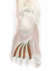
The four lumbrical muscles of the foot are small skeletal muscles on the foot's plantar surface, extending the lateral toes from the interphalangeal joints and flexing them from the metatarsophalangeal joints. Numbered one through four from the medial side outward, the first and most medial of the lumbrical muscles is somewhat distinct from the other three more lateral muscles, being served by different nerves and arteries among other distinctions. Innervated by the medial plantar nerve (on the most medial of the lumbrical foot muscles) and lateral plantar nerve branches of the tibial nerve, the lumbrical muscles help articulate and curl the lesser toes. They receive their blood supply from the medial and lateral plantar arteries. The lumbricals serve as accessory muscles to the flexor digitorum longus, which runs across the sole of the foot and the medial (tibial) side of the heel. The origin of the first lumbrical muscle is a single head on the medial side of the flexor digitorum longus, while the other three lumbricals each arise from two heads between their respective tendons. All four lumbrical muscles pass along the medial sides of their respective toes to insert into extensor tendons of the extensor digitorum longus on the front of the foot at the proximal phalanges.

Copyright © Innerbody Research 1997 - 2025. All Rights Reserved.
