The Muscles of the Eye
Explore the anatomy and function of the eye muscles with Innerbody's interactive 3D model.

Six skeletal muscles surround the eye and control the many diverse movements of the eyes. These muscles, although small and not particularly strong, are exceptionally fast and precise. They allow the eye to perform many complex tasks, including tracking moving objects, scanning for objects, and maintaining a stable image on the retina.
Anatomy
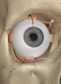
Six skeletal muscles surround and move the eye, working against each other to produce various eye movements. All of these muscles, except for the inferior oblique muscle, arise from a tendinous band surrounding the optic nerve. This band, known as the annulus of Zinn, anchors the muscles of the eye to the posterior of the orbit while providing holes for the nerves of the eye to enter the orbit.
The superior rectus muscle forms its origin on the annulus of Zinn and inserts along the superior edge of the eye. It is a thin muscle and forms a straight muscular band between the eye and the annulus of Zinn. The inferior, medial, and lateral rectus muscles are almost identical to the superior rectus muscle, except they insert on the inferior, medial, and lateral edges, respectively, of the eye.
Compared to the four rectus muscles of the eye, the two oblique muscles follow unique paths and insert on the eye at oblique angles. Arising from the annulus of Zinn, the superior oblique muscle passes through a ring of connective tissue known as the trochlea before inserting obliquely on the superior surface of the eye. The trochlea is located on the roof of the orbit medial and anterior to the eye and redirects the tendon of the superior oblique muscle to reach the eye from the anterior and medial direction. Unique among the muscles of the eye, the inferior oblique muscle arises from the maxilla on the medial floor of the orbit and inserts at an oblique angle on the inferior surface of the eye.
Physiology
The four rectus muscles of the eye control the movement of the eye in the cardinal directions. They work against each other to control the movements of the eye in various directions. The first of these muscles, the superior rectus muscle, elevates the eye, allowing the eye to look up. The antagonist of the superior rectus muscle is the inferior rectus muscle, which depresses the eye, allowing the eye to look down. On the medial side of the eye, the medial rectus muscle adducts the eye, allowing it to look medially towards the nose. Its antagonist is the lateral rectus muscle that abducts the eye, allowing it to look laterally or away from the body's midline.
The two oblique muscles of the eye are responsible for the rotation of the eye and assist the rectus muscles in their movements. The superior oblique muscle rotates the eye medially and abducts it when the eye if facing forward while the inferior oblique rotates the eye laterally and adducts it. When the eye is adducted, or turned toward the nose, the superior oblique depresses the eye while the inferior oblique elevates the eye.
The muscles of the eyes perform several specialized functions to aid in vision. When looking at a large area, the muscles perform a scanning function, known as saccades, to provide vital information to the brain. During saccades, the eyes dart between several points in the field of view to provide information about the scene to the brain. A small region of the retina, known as the fovea, has the highest concentration of cones and produces the most detailed visual images. Saccades allows the fovea to produce clear images of the most important parts of an image to the brain for fast analysis.
Another important function is tracking moving objects across the visual field. The brain sends many signals to the muscles of the eye to quickly adjust the eye's position so that a moving object of interest remains within the fovea and can be seen clearly.
Finally, the muscles of the eye adjust the position of each eye subtly to produce a single, binocular image. This process is known as vergence, and prevents the double vision and blurred vision that would result from different images being presented to the visual cortex of the brain. Looking down at the tip of your nose or crossing your eyes voluntarily stops vergence, producing double vision and an uncomfortable sensation.

Copyright © Innerbody Research 1997 - 2025. All Rights Reserved.
