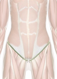
The Inguinal Ligament
Explore the anatomy and important function of the inguinal ligament in the groin region with Innerbody's 3D model.

The inguinal ligament is an important connective tissue structure in the inguinal, or groin, region of the human body. It supports soft tissues in the groin as well as the external abdominal oblique muscle.
The inguinal ligament is a narrow band of dense regular fibrous connective tissue in the pelvic region of the body. Its collagen fibers arise from the inferior aponeurosis of the external abdominal oblique and run obliquely across the pelvis. On its superior and lateral end it connects to the anterior iliac spine of the ilium and extends to the pubic tubercle of the pubis bone on its inferior and medial end.
The inguinal ligament supports the muscles that run inferior to its fibers, including the iliopsoas and pectineus muscles of the hip. It also supports the nerves and blood vessels of the leg as they pass through the groin, including the femoral artery, femoral vein, and femoral nerve. The support provided by the inguinal ligament is important to maintaining the flexibility of the hip region while allowing vital blood and nerve supply to the leg.
A small opening in the muscles and connective tissues of the abdomen --- known as the superficial inguinal ring --- is located just superior to the inguinal ligament. This opening forms part of the inguinal canal, which permits the spermatic cord in males and the round ligament of the uterus in females to exit the abdominopelvic cavity and pass through the external tissues of the pelvis. The inguinal ligament forms the floor of the inguinal canal and supports the passage of structures through the canal.
Inguinal hernias are a complication of the anatomical arrangement of the inguinal canal, especially in males. The spermatic cord requires that the inguinal canal remain open to permit the passage of blood vessels, nerves, and the ductus deferens into the scrotum. Under strenuous contraction of the abdominal muscles, the pressure on the organs of the abdominopelvic cavity can become so great that a segment of the small intestine can be forced through the inguinal canal, resulting in an inguinal hernia. Surgery for this condition may include the grafting of reinforcing mesh material to the inguinal ligament to prevent further herniation.


