Ureter

The ureters are a pair of small tubes that connect the kidneys to the urinary bladder. They form a vital link in the urinary tract by allowing urine to drain from the kidneys to be stored in the bladder.
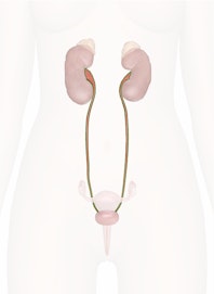
Anatomy
The ureters are thin, muscular tubes found within the abdominopelvic cavity. Each is about 10 to 12 inches (25-30 cm) long, with the left ureter being slightly longer than the right due to the left kidney lying superior to the right kidney. The width of the ureters is only a few millimeters in diameter and varies from one end to the other --- wider near the kidneys and narrower near the urinary bladder.
Each ureter begins at the kidney where the renal pelvis exits through the renal hilus. The ureter begins as a tube about 10 mm in diameter as it exits the kidney, but it tapers to only 1-2 mm as it descends through the abdominopelvic cavity. Like the kidneys, the ureters are retroperitoneal organs, which pass through the walls of the abdominopelvic cavity posterior to the peritoneum. Initially the ureters point medially, but quickly turn inferiorly and descend toward the pelvis. Within the pelvis, the ureters turn anteriorly to pass over the common iliac arteries and veins before turning posteriorly to resume their original courses. At the base of the pelvis, the ureters once again turn anteriorly to approach the urinary bladder and enter the bladder on its posterior wall. The ureters pass obliquely through the wall of the urinary bladder and end at the ureteral openings about 1 inch (2 cm) within the bladder.
Histology
The wall of the ureter is made of several distinct tissue layers. The innermost layer of the ureter is made of mucosa featuring transitional epithelium that is able to stretch to accommodate varying volumes of urine and prevent wastes from escaping into the body. Surrounding the mucosa is the submucosa layer that contains the connective tissue, blood vessels, and nerves that support the other layers of the ureter. A thick layer of smooth muscle tissue, known as the muscularis, surrounds the submucosa, provides the ureter with the ability move, and adjusts its aperture. Finally, the outermost layer of the ureter, the adventitia, is made of loose connective tissue that loosely anchors the ureter to the surrounding tissue while providing some freedom of movement.
Physiology
While the ureters may appear to be simple tubes for urine to drain into the urinary bladder, they actually play active roles in the urinary system. Under most circumstances, gravity is able to pull urine down from the kidneys to the urinary bladder. However, gravity would not be able to move urine to the bladder when one is lying down or in freefall (such as an astronaut in space). To ensure the unidirectional flow of urine, the ureters use waves of smooth muscle contraction, known as peristalsis, to conduct urine to the urinary bladder. Once urine has entered the urinary bladder, the pressure of the inflated bladder squeezes the ends of the ureters, preventing urine from flowing back towards the kidneys. These processes for keeping urine flowing away from the kidneys help to eliminate waste in the most efficient way possible and help to prevent bacterial infections from reaching the kidneys.

Copyright © Innerbody Research 1997 - 2025. All Rights Reserved.
