Eye

The eye is one of the most complex and important sensory organs in the human body. It provides the ability to see in both bright and dim light, focusing on objects both near and far. Its three types of cone cells are able to distinguish millions of distinct colors and produce the high quality images that the body relies on for many daily activities.
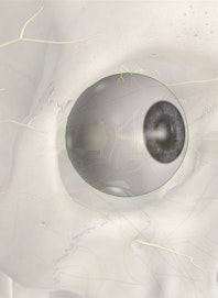
Eye Anatomy
The eyes are a pair of spherical organs located within the orbits of the skull. Each eye is roughly 1 inch (2.5 cm) in diameter and fills most of the space within the orbit. Three distinct tissue layers --- the fibrous, vascular, and nervous tunics --- make up the wall of the eye and surround its gel-filled center.
The fibrous tunic forms the outermost layer of the eye and is instrumental in protecting its delicate inner tissues.
- Anteriorly, the fibrous tunic consists of the cornea, a clear layer of dense regular fibrous connective tissue. The cornea forms a circular window allowing light to enter the eye while blocking foreign material from entering.
- Extending from the cornea to cover the sides and posterior of the eye is the sclera. The sclera is a thick, white layer of dense irregular connective tissue that acts like a tough yet flexible shell for the eye. A thin layer of mucous membrane known as the conjunctiva covers the anterior surface of the sclera and the inside of the eyelids. The conjunctiva secretes mucus to lubricate the surface of the eye and contains blood vessels that support the tissues of the sclera.
Deep to the fibrous tunic is the vascular tunic that provides blood supply to the eye. It has three major parts: the choroid, ciliary body, and iris.
- The choroid is a layer of connective tissues lining the inside of the sclera and providing blood flow to the sclera and retina. It also contains a high concentration of melanin, giving it a black color and helping it to absorb light in the eye.
- The ciliary body is a widened ring of tissue at the anterior edge of the choroid. It contains the ciliary muscles and the ciliary bodies. The ciliary muscles pull on the zonular fibers and the lens of the eye to focus light, while the ciliary bodies produce aqueous humor.
- The iris is a ring of pigmented smooth muscle extending from the anterior edge of the ciliary body and surrounding the pupil. Movement of the smooth muscle tissue in the iris adjusts the size of the pupil, the circular hole in the center of the iris.
The lens is a large, clear mass of protein fibers found just posterior to the iris. It is connected to the ciliary body by many tiny fibers known as zonular fibers. The lens is extremely flexible and changes its shape when the ciliary body pulls on its edges.
The innermost layer of the eye is formed by the nervous tunic, or retina. It is a thin, delicate layer of nervous tissue that is loosely connected to the choroid. Millions of photoreceptor cells, bipolar cells, and ganglion cells are distributed throughout the retina to detect light and transmit visual information to the brain. The macula lutea is the region of central vision, which contains the fovea. The fovea is an important region of the retina, as it contains the highest concentration of photoreceptors in the eye. Nervous tissue in the retina is connected to the brain through the optic nerve, which passes through a hole in the choroid and sclera in the posterior of the eye. Where the optic nerve forms there are no photoreceptors, resulting in a blind spot known as the optic disk.
The lens divides the interior of the eye into two regions: the anterior chamber and the vitreous chamber.
- Posterior to the lens is the vitreous chamber, which is much larger than the anterior chamber. The vitreous humor, a clear, gel-like mass of water and proteins, fills the vitreous chamber. It gives shape to the eye, holds the retina in place against the choroid, and allows light to pass easily through the eye.
- The anterior chamber is filled with aqueous humor, a clear liquid produced by the ciliary body. Aqueous humor plays a number of roles in the eye, including inflating the eye through intraocular pressure; providing a clear medium for light to pass through the eye; and nourishing the cells of the cornea and lens.
Physiology of the Eye
The overall function of the eye is to act like a biological camera---it absorbs light and translates images into nerve signals to conduct to the brain. Light entering the eye first passes through the cornea, where it is refracted to begin the process of focusing. It next passes through the pupil, where the contraction of muscles in the iris controls the size of the pupil and the amount of light entering the eye. Light passes through the lens, where it is further refracted to focus on the retina. Contractions of the ciliary muscles pull on the zonular fibers and the lens, allowing the lens to accommodate vision at varying differences. To view objects that are close to the eye, the ciliary muscles relax and permit the lens to assume a wide shape. The wide shape of the lens allows it to refract the light to a high degree to focus on the retina. For distant objects, the ciliary muscles pull on the lens to flatten it, reducing the amount of refraction and focusing distant light on the retina.
Once light has passed through the lens, it continues through the vitreous humor and passes through the retina. Photoreceptor cells in the retina are specialized to detect light and produce nerve signals in response to light. Rods are the more numerous and sensitive of the photoreceptors and are specialized for seeing in low-light situations. They produce grayscale images in low light, but are overwhelmed by light during the day or in a normally lit room at night. Cones, on the other hand, are specialized for detecting light in brighter conditions and are able to differentiate colors. The three types of cone cells --- red, green, and blue --- are able to detect specific colors, or wavelengths, of light. The combination of the three types of cone cells produces all of the colors that the human eye can detect. Once the photoreceptors have detected light, the cells produce an action potential that is conducted to bipolar cells and ganglion cells in the retina. These cells transmit the signal into the optic nerve, where it travels to the brain to be processed.
After light has passed through the retina, it is absorbed by the choroid. The choroid prevents excess light from remaining within the eye and forming afterimages. High intensity lights can overcome the absorptive effects of the choroid, resulting in the "red eye" seen in pictures.
Health Concerns
Our eyes are susceptible to many health problems---astigmatism, infection, nearsightedness, cataracts, glaucoma and macular degeneration, to name a few. Learn more about how DNA health tests can tell you if you have a genetically higher risk of developing macular degeneration.
READ THIS NEXT


-new-upper.jpg?auto=format,compress&fit=max&w=262&q=75)


Copyright © Innerbody Research 1997 - 2025. All Rights Reserved.
