Intercalated Discs: Heart Cell Connection & Signal Transmission
Discover the intercalated discs, structures in the heart, and components including fascia adherens and gap junctions. Learn about their roles in cardiac function.

Intercalated discs are unique structural formations found between the myocardial cells of the heart. They play vital roles in bonding cardiac muscle cells together and in transmitting signals between cells.
Anatomy
The ends of each cardiac muscle cell form intercalated discs where they meet neighboring cardiac muscle cells. Each intercalated disc contains many finger-like extensions of plasma membrane that interlock with identical structures on the neighboring cell. The structure of the membrane in the intercalated disc greatly increases the surface area contact between the cells and helps to hold the cells together.
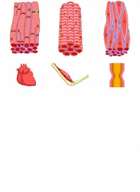
Three types of special structures --- fascia adherens, desmosomes, and gap junctions --- are found in the intercalated discs to help in the connection of the cardiac muscle cells.
- The fascia adherens are bands of proteins that connect the actin filaments of the sarcomeres in each cardiac muscle fiber to the sarcomere in the neighboring cells, producing a single unified chain of sarcomeres.
- Desmosomes help to bind cardiac muscle cells together, but form smaller, tighter junctions compared to the fascia adherens. Intermediate fibers inside each muscle fiber are connected by a series of proteins in the desmosome, which form an interlocking protein chain.
- Finally, many gap junctions form between the cardiac muscle cells to allow material to pass between the cells. Each gap junction is made of connexin proteins that form a tunnel through the cell membranes of the cardiac muscle cells, allowing small molecules, including ions, to pass through.
Physiology
Cardiac muscle cells produce strong contractions to pump blood throughout the body, but these same contractions threaten to destroy the structures of the heart. Intercalated discs play a vital role in holding cardiac muscle cells together using the fascia adherens and desmosomes. The increased surface area of the finger-like extensions of cell membrane provides ample room for these connections to form. Gap junctions allow ions to pass from one cardiac muscle cell to another, providing an extremely fast conduction of action potentials through cardiac muscle tissue. The net result of the strong connections and fast ion conduction is the contraction of cardiac muscle tissue like a single functional unit.

Copyright © Innerbody Research 1997 - 2025. All Rights Reserved.
