The Terminal Ileum
Explore the anatomy and role of the terminal ileum with Innerbody's interactive 3D model.

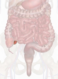
The terminal ileum is the distal end of the small intestine that intersects with the large intestine. It contains the ileocecal sphincter, a smooth muscle sphincter that controls the flow of chyme into the large intestine.
Anatomy
The terminal ileum is located on the right side of the abdominopelvic cavity in the umbilical and hypogastric regions. It is a tube about 1.25 to 1.5 inches (3 to 4 cm) long at the end of the ileum and terminates at the ileocecal sphincter. The ileocecal sphincter is a band of smooth muscle that controls the flow of chyme from the ileum into the cecum of the large intestine.
Histology
The walls of the terminal ileum are made of four distinct tissue layers surrounding the hollow lumen at its center.
- The innermost layer, the mucosa, contains epithelial cells whose function is specialized for the absorption of nutrients from chyme in the lumen. Masses of lymphatic tissue known as Peyer's patches dot the surface of the mucosa to detect the presence of pathogens in the chyme.
- Surrounding the mucosa is the submucosa layer that contains the nerves and blood vessels, which support the other tissue layers of the terminal ileum.
- The muscularis layer surrounds the submucosa and contains bands of smooth muscle that contract in wave of peristalsis to move food through the terminal ileum and into the cecum.
- Finally, the serosa forms the outermost layer of the terminal ileum. Serosa is made of a smooth layer of simple squamous epithelial tissue and secretes a slippery liquid known as serous fluid. Serosa and serous fluid give the terminal ileum a slick, slimy surface to protect it from friction between organs and the walls of the abdominopelvic cavity.
Physiology
Chyme is thoroughly processed by the duodenum, jejunum, and ileum before it enters the terminal ileum. It is then stored in the hollow lumen of the terminal ileum as it awaits the opening of the ileocecal sphincter. Small masses of chyme are pushed into the cecum by waves of peristaltic contraction of the walls of the terminal ileum coordinated with the opening of the ileocecal sphincter. While the chyme is stored, Peyer's patches lining the walls of the terminal ileum examine the contents of the chyme for any potentially dangerous pathogens.

Copyright © Innerbody Research 1997 - 2025. All Rights Reserved.
