The Chordae Tendineae
Discover the anatomy and function of the chordae tendineae with Innerbody's interactive 3D model.

The chordae tendineae are a group of tough, tendinous strands in the heart. They are commonly referred to as the "heart strings" since they resemble small pieces of string. Functionally, the chordae tendineae play a vital role in holding the atrioventricular valves in place while the heart is pumping blood.
Anatomy
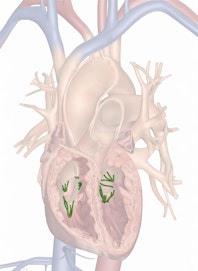
The chordae tendineae are a group of string-like tendinous bands found within both ventricles of the heart. They arise from the tips of the papillary muscles on the inside of the wall of the ventricles and extend into the hollow lumen. Most separate into two or more branches, but some resemble simple, unbranched strings. On their far end, the chordae tendineae merge with and insert on the cusps of the atrioventricular (AV) valves. In the right ventricle, the chordae tendineae connect to the three cusps of the tricuspid valve, while in the left ventricle they connect to the two cusps of the bicuspid (or mitral) valve.
Histologically, the chordae tendineae resemble thin tendons made of dense regular connective tissue. Most of their mass is made of strong collagen protein fibers, but they also contain elastin protein fibers to provide elasticity and fibroblast cells to repair the protein fibers.
Physiology
The purpose of ventricular systole, or contraction of the ventricles, is to pump blood out of the heart and into the aorta and pulmonary trunk. However, blood will exit the ventricles through any opening, and some blood attempts to regurgitate, or flow backwards, into the atria. The AV valves prevent regurgitation by covering the openings to the atria and forcing blood to exit the heart. To prevent the valves from blowing out under the extremely high blood pressure within the ventricles, the chordae tendineae hold the cusps of each AV valve on the ventricular side. Once filled with blood, the AV valves form a dome shape and resemble tiny parachutes with the chordae tendineae acting as the strings.

Copyright © Innerbody Research 1997 - 2025. All Rights Reserved.
