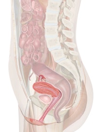
![]() |
||||||||||||||||||||||||||||||||||||||||||||||||||||||||||||||||
|
|
AnusThe anus is a canal at the end of the digestive tract through which feces is expelled. It is about five inches long and is an extension of the rectum. It is only open during the expulsion of feces, because it is usually kept closed by sphincter muscles, which can be relaxed at will. ClitorisThe clitoris is a small projection at the front of the vulva between the labia minor. Although most of it is embedded in surrounding tissues, it is usually about 2 cm long and 0.5 cm in diameter. The clitoris corresponds to the penis in the male and is somewhat similar in structure. It is composed of two columns of erectile tissue, which are separated by a septum and are surrounded by a covering of dense, fibrous connective tissue. At the root of the clitoris, the columns of tissue diverge to form "crura," which in turn are attached to the sides of the pubic arch. At the front, a small mass of erectile tissue forms a "glans," which is richly supplied with sensory nerve fibers. Labia MajorThe labia major (singular, labium) enclose and protect the other external reproductive organs. They correspond to the scrotum of the male and are composed primarily of rounded folds of adipose tissue covered by skin. On the outside the skin includes numerous hairs, sweat glands, andsebaceous glands, while on the inside it is thinner and hairless. The labia major lie closely together and are separated lengthwise by a cleft that includes the urethral and vaginal openings. At the front, the labia merge to form a rounded elevation of fatty tissue called the "mons pubis," which overlies the symphysis pubis. At the back, near the anus, the labia are somewhat tapered and merge into the labia minor. Labia MinorThe labia (singular, labium) minor are flattened lengthwise into folds located with the cleft between the labia major. These folds extend along either side of the vestibule. They are composed of connective tissue that is richly supplied with blood vessels, causing a pinkish appearance. In the back, near the anus, the labia minor merge with the labia major, while in the front they converge to form a hood-like covering around the clitoris. Pubic SymphysisThe pubic symphysis is a cartilage-like articulation between the pubic bones. It is a slightly movable joint that contains three ligaments: the superior pubic, the arcuate pubic and the interpubic disk. RectumThe rectum is a short, muscular tube that forms the lowest portion of the large intestine and connects it to the anus. Feces collects here until pressure on the rectal walls cause nerve impulses to pass to the brain, which then sends messages to the voluntary muscles in the anus to relax, permitting expulsion. SacrumThe sacrum is a large triangular bone at the base of the lower spine. Its broad upper part joins the lowest lumbar vertebrae and its narrow lower part joins the coccyx or "tail bone". The sides are connected to the iliums (the largest bones forming the pelvis). The sacrum is a strong bone and rarely fractures. The five vertebrae that make up the sacrum are separated in early life, but gradually become fused together between the eighteenth and thirtieth years. The spinous processes of these fused bones are represented by a ridge of tubercles. The sacrum is wedged between the coxal bones of the pelvis and is united to them by fibrocartilage at the sacroiliac joints. The weight of the body is transmitted to the legs through the pelvic girdle at these joints. SkinSkin is the outside covering of body tissue, which protects inner cells and organs from the outside environment. The skin is the largest organ of the body, and its cells are continuously replaced as they are lost to normal wear and tear. The skin totals between twelve and twenty square feet in area and accounts for 12%% of body weight. It is composed of three integrated layers: the epidermis, the dermis and the subcutis. The thickness of the epidermis and the dermis varies over different parts of the body. It is thickest on the palms of the hands and feet, where friction is needed for gripping, and it is thinnest on the eyelids, which must be light and flexible. The epidermis also grows into fingernails, toenails and hair. The dermis, or true skin, is thick, sturdy, rich in nerves and blood vessels and in sweat glands. It shields and repairs injured tissue. This layer consists mostly of collagen, which originates from cells called fibroblasts and is one of the strongest proteins found in nature. It gives skin durability and resilence. The subcutis, joined to the bottom of the dermis, is the deepest layer of the skin. It contains "lipocytes," which produce lipids for the subcutaneous tissue to make a fatty layer which cushions muscles, bones and inner organs against shocks, and acts as an insulator and source of energy during lean times. The skin registers sensation constantly and supports a teeming, unseen population of tiny organisms. Not only does the skin harden from use, but it molds into varied shapes, and it responds to the most delicate touch, becoming an organ of communication - sometimes more eloquent than words. So tough and durable is the skin that when a 2,000-year-old Egyptian mummy was fingerprinted, the ridges were perfectly preserved. Small IntestineIf the small intestine were not looped back and forth upon itself, it could not fit into the abdominal space it occupies. It is held in place by tissues which are attached to the abdominal wall and measures eighteen to twenty-three feet in the average adult, which makes it about four times longer than the person is tall. It is a three-part tube of about one and one-half to two inches in diameter and is divided into three sections: (1) the duodenum, a receiving area for chemicals and partially digested food from the stomach; (2) the jejunum, where most of the nutrients are absorbed into the blood and (3) the ileum, where the remaining nutrients are absorbed before moving into the large intestine. The intestines process about 2.5 gallons of food, liquids and bodily waste every day. In order for enough nutrients to be absorbed into the body, it must come in contact with large numbers of intestinal cells which are folded like gathered skirts. Each of these cells contain thousands of tiny finger-like projections called "villi," and each villus contains microscopic "microvilli". In one square inch of small intestine, there are about 20,000 villi and ten billion microvilli. Each villus brings in fresh, oxygenated blood and sends out nutrient-enriched blood. The villi sway constantly to stir up liquefied food and remove the nutrients which can be absorbed and then passed through the membranes of the villi into the blood and lymph vessels. The fatty nutrients go to the lymph vessels, and glucose and amino acids go to the blood and on to the liver. The muscles which encircle this tube constrict about seven to twelve times a minute to move the food back and forth, to churn it, knead it, and to mix it with gastric juices. The small intestine also makes waves which move the food forward, but these are usually weak and infrequent to allow the food to stay in one place until the nutrients can be absorbed. If a toxic substance enters the small intestine, these movements may be strong and rapid to expel the poisons quickly. Sphincter MusclesA "sphincter" is a ring of muscle around a natural opening or passage that acts like a valve, regulating inflow and outflow. The outlet from the stomach into the duodenum is the "pyloric" sphincter and controls the stomach's outflow. The "anal" sphincter alters rectal outflow and is partly voluntary, allowing a decision on when to empty the bowel. UrethraThe urethra is a tube that conveys urine from the urinary bladder to the outside of the body. Its wall is lined with mucous membranes and contains a relatively thick layer of smooth muscle tissue. It also contains numerous mucous glands, called "urethral glands," that secrete mucus into the urethral canal. In females the urethra is about 4 cm long. It passes forward from the bladder, descends below the symphysis pubis, and empties into the labia minor. Its opening is located above the vaginal opening and about 2.5 cm below the clitoris. In males, the urethra, which functions both as a urinary canal and a passageway for cells and secretions from various reproductive organs, can be divided into three sections: the prostatic urethra, the membranous urethra, and the penile urethra. Urinary BladderThe bladder is a hollow muscular organ that acts as a reservoir for urine. The adult bladder can hold a pint or more of fluid. It lies behind the pubic bone and is protected by the pelvis. The bladder walls are made up of muscle and an inner lining. At the back are two ureters which carry the urine from the kidneys. The bladder collects and stores urine until it can be expelled from the body. Urogenital DiaphragmThe outlet of the pelvis is spanned by two muscular sheets - a deeper "pelvic diaphragm" and a more superficial "urogenital diaphragm." The pelvic diaphragm forms the floor of the pelvic cavity, and the urogenital diaphragm fills the space within the pubic arch. The muscles of the male and female urogenital diaphragm include the following: (1) "superficial transversus perinei," a small bundle of muscle fibers that pass along the back border of the urogenital diaphragm and assists other muscles in supporting the pelvic tissues; (2) the "bulbospongiosus" muscles, which are united and surround the base of the penis; (3) the "ischiocavernosus muscle," a tendinous structure that extends down to the margin of the pubic arch and assists the function of #2; and (4) the "sphincter urethrae" are muscles that arch around the urethra and unite with those on the other side. Together they act as a sphincter that closes the urethra by compression and opens it by relaxation to control the flow of urine. UterusThe uterus or "womb" is a hollow, muscular organ in which a fertilized egg, called the "zygote," becomes embedded and in which the egg is nourished and allowed to develop until birth. It lies in the pelvic cavity behind the bladder and in front of the bowel. The uterus usually tilts forward at a ninety degree angle to the vagina, although in about 20% of women, it tilts backwards. The uterus is lined with tissues which change during the menstrual cycle. These tissues build under the influence of hormones from the ovary. When the hormones withdraw after the menstrual cycle, the blood supply is cut off and the tissues and unfertilized egg are shed as waste. The endometrium is the membrane that lines the uterus. It is a glandualr mucous membrane, and is formed during the menstrual cycle, then expelled from the body during each menstruation. This occurs in females on average every twenty-eight days, but varies with each person. As the endometrium is expelled, the blood vessels which connect the endometrial lining to the uterus break, causing bleeding and usually a small degree of pain. The endometrium is what a fertilized egg attaches to upon fertilization, so it is not shed if a fertilized egg is implanted. The endometrium also supplies blood for the fetus and allows it to receive oxygen. During pregnancy, the uterus stretches from three to four inches in length to a size which will accommodate a growing baby. During this time, muscular walls increase from two to three ounces to about two pounds and these powerful muscles release the baby through the birth canal with great force. The womb shrinks back to half its pregnant weight before a baby is a week old. By the time the baby is a month old, the uterus may be as small as when the egg first entered. Superstition, myth or ignorance have surrounded the menstrual period since the beginning of time. This is largely due to a primitive fear of blood. The word, "taboo," may stem from the Polynesian word for menstruation, but not all legends are negative; a girl's first menses is celebrated in some societies, because it is a sign that she can bear children. VaginaThe vagina is a muscular passage which forms a part of the female sex organs and which connects the neck of the uterus (called the "cervix") with the external genitals. The vagina, which is approximately two and one-half to four inches long, has muscular walls which are supplied with numerous blood vessels. These walls become erect when a woman is aroused as extra blood is pumped into these vessels. The vagina has three functions: as a receptacle for the penis during love-making; as a outlet for blood during menstruation; and as a passageway for the baby to pass through at birth. According to The Guiness Book of World Records, a Russian peasant woman who lived in the 18th Century holds the record for the most children born to one mother. She had sixty-nine children within forty years. She produced sixteen pairs of twins, seven sets of triplets, and four sets of quadruplets! |
|||||||||||||||||||||||||||||||||||||||||||||||||||||||||||||||