The Larynx
Explore the anatomy and role of the larynx in the respiratory tract with Innerbody's 3D model.

The larynx is a tough, flexible segment of the respiratory tract connecting the pharynx to the trachea in the neck. It plays a vital role in the respiratory tract by allowing air to pass through it while keeping food and drink from blocking the airway. The larynx is also the body's "voice box" as it contains the vocal folds that produce the sounds of speech and singing.
Anatomy
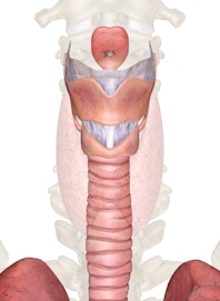
The larynx is a short, epithelium-lined tube formed by nine pieces of cartilage and several ligaments that bind them together. It is located along the body's midline in the neck region deep to the skin and the muscles of the neck and anterior to the esophagus and cervical vertebrae. At its superior end, it borders the hyoid bone and the laryngopharynx.
The most superior region of the larynx is the epiglottis, a leaf-shaped flap of elastic cartilage covered with epithelium. It connects to the larynx on its tapered inferior end and, except for a brief moment while swallowing, extends its wider superior end slightly into the pharynx just posterior to the tongue. During the process of swallowing, the epiglottis folds over to cover the glottis and prevents food from blocking the airway.
Inferior to the epiglottis is the glottis region of the larynx, which contains the vocal folds. The largest cartilage in the larynx, the thyroid cartilage, supports the glottis. The thyroid cartilage is semicircular in shape with a prominent ridge extending from its anterior surface. This ridge is larger in males than in females and is visible through the skin of the neck, forming the structure known as the Adam's apple. The thyroid cartilage is connected on its superior surface to the hyoid bone by a wide ligament known as the thyrohyoid membrane. The thyroid cartilage also anchors the anterior ends of the vocal folds, which attach to the inside of the thyroid cartilage at the body's midline.
The cricoid cartilage is the most inferior structure of the larynx and forms the transition between the larynx and the trachea. It is ring-shaped, with its widest portion facing posteriorly and its narrowest portion facing anteriorly. The cricothyroid ligament connects the cricoid cartilage to the thyroid cartilage along most of its superior surface, while the cricotracheal ligament connects it to the trachea along its inferior surface. The wide posterior of the cricoid cartilage almost touches the thyroid cartilage and forms the cricothyroid joint. Sound pitch is modified by adjustment of the angle of the cricothyroid joint, which helps to control the tension of the vocal folds.
Posterior to the thyroid cartilage are three tiny, paired masses of cartilage known as the cuneiform, corniculate, and arytenoid cartilages. The arytenoid cartilages are roughly pyramidal masses of cartilage that sit on top of the posterior side of the cricoid cartilage. These tiny cartilages play a vital role in the production of sound and act as the pivot points for the vocal folds and the muscles that move the vocal folds. Resting on top of the arytenoid cartilages are the horn-shaped corniculate cartilages that help to support the posterior end of the glottis. The cuneiform cartilages are long, thin bands of cartilage that extend laterally from the corniculate cartilages and support the lateral sides of the glottis.
Physiology
In the process of swallowing, the larynx plays an important role in the direction of food into the esophagus. The epiglottis normally resides in an upright position just anterior to the lumen of the larynx. In this position, it allows air to pass freely through the larynx during inhalation and exhalation. When food or liquid in the mouth is swallowed, the food pushes the epiglottis posteriorly, flipping its free edge over to cover the glottis and block the swallowed substances from entering the larynx. The food then safely passes on to the esophagus, at which point the epiglottis flips back to its resting position.
Occasionally, a person may choke when food gets past the epiglottis or sticks to a structure within the pharynx and blocks the airway. The vocal folds contract to catch the blockage before it passes into the trachea. Coughing pushes air out of the lungs to force the blockage out of the airway.
Sounds are produced in the larynx by the movement of air through the larynx and by the vocal folds, a pair of movable folds in the mucous membrane. The vocal folds are connected to the thyroid cartilage on their anterior ends and the arytenoid cartilages on their posterior end. Air exhaled from the lungs passes through the larynx and vibrates the vocal folds. Several sets of muscles move the arytenoid cartilages and the cricothyroid joint to adjust the position and tension of the vocal folds and thereby control the pitch of sound made by the larynx.
READ THIS NEXT


Copyright © Innerbody Research 1997 - 2025. All Rights Reserved.
