Otic Ganglion

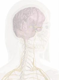
The otic ganglion is shaped like an oval. It's a reddish-gray color and is a small, flat ganglion that is positioned right under the foramen ovale. It rests on the mandibular nerve's medial surface and borders the place where the nerve to the pterygoideus internus originates. The otic ganglion runs parallel to the mandibular nerve's trunk where the sensory and motor roots connect. It flows in the middle of the tensor veli palatini's origin and the cartilaginous part of the auditory tube. It runs in posteriorly with the middle meningeal artery.
The middle meningeal artery's plexus is where the otic ganglion's sympathetic root appears. The inferior maxillary creates its sensory root by way of the internal pterygoid nerve. The small superficial petrosal nerve that sends and receives information with the tympanic branch of the glossopharyngeal nerve is where the motor root comes into existence. The otic ganglion exchanges information with the auriculotemporal and chorda tympani nerves. The inferior maxillary nerve's motor fibers run along the otic ganglion to reach the tensor palate and tympani tensor muscles.

Copyright © Innerbody Research 1997 - 2025. All Rights Reserved.
