Nail Body of Toenail

The nail body of the toenail is the principle portion of the human nail. The nail body, or nail plate, is made of keratin that is translucent. It is the exposed portion of the nail, growing from the nail matrix, beyond the nail root, and out from beneath the cuticle or eponychium. Laterally, the nail body is enclosed within the nail groove by the flexible tissue of the paronychium, and it terminates at the distal edge or free margin of the nail, which hangs free of the nail bed. It looks pink because of the blood vessels located under the nail, although there are no blood vessels located in the nail plate. It is porous to water and the amount of water it holds is connected to how brittle and hard it is. The nail plate's calcium content is less than 0.5%.
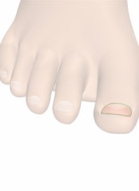
At the junction of the nail body proper and the distal edge of the nail is the hyponychium, a protective layer of epithelial cells that seals the end of the nail bed and epidermis to the nail at the onychodermal band. While the nail is constantly growing, the nail body is nevertheless firmly attached to the skin of the nail bed beneath it. The layered cells of the nail body are keratin cells, like those of the hair, flattened into a thin plate with longitudinal grooves that may be barely visible. By the time they are exposed to the air beyond the cuticle, these cells are dead. The growing and dividing cells of the nail matrix and nail root continually push the dead keratinized epithelial cells of the nail body forward.

Copyright © Innerbody Research 1997 - 2025. All Rights Reserved.
