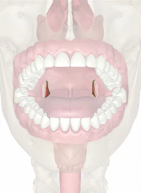
Palatopharyngeal Arch and Palatopharyngeus Muscle

The palatopharyngeus muscle, also known as the pharyngopalatinus muscle, is a long bundle of tissue. When these muscles contract, they pull the pharynx up and cover the bolus of food. They almost meet, but the uvula fills up the small space between them. These actions prevent the bolus from moving along into the nasopharynx. These muscles also make up an inclined plane. This plane is positioned down and back, on the underside of the surface where the bolus passes downward into the pharynx's lower part. It is thicker at the ends than it is in the middle; along with the mucous membrane, which covers its entire surface, this muscle forms the palatopharyngeal arch.
This arch is the last of the two mucous membrane folds on either side of the oral part of the pharynx (which extends from the uvula to the hyoid bone). Its purpose is to surround the palatopharyngeus muscle and be the fauces' posterior pillar.

Copyright © Innerbody Research 1997 - 2025. All Rights Reserved.
