Respiratory System (Male Posterior View)

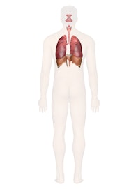
The cells of the human body require a constant stream of oxygen to stay alive. The respiratory system provides oxygen to the body's cells while removing carbon dioxide, a waste product that can be lethal if allowed to accumulate. There are 3 major parts of the respiratory system: the airway, the lungs, and the muscles of respiration. The airway, which includes the nose, mouth, pharynx, larynx, trachea, bronchi, and bronchioles, carries air between the lungs and the body's exterior. The lungs act as the functional units of the respiratory system by passing oxygen into the body and carbon dioxide out of the body. Finally, the muscles of respiration, including the diaphragm and intercostal muscles, work together to act as a pump, pushing air into and out of the lungs during breathing.
Anatomy of the Respiratory System
Nose and Nasal Cavity
The nose and nasal cavity form the main external opening for the respiratory system and are the first section of the body's airway---the respiratory tract through which air moves. The nose is a structure of the face made of cartilage, bone, muscle, and skin that supports and protects the anterior portion of the nasal cavity. The nasal cavity is a hollow space within the nose and skull that is lined with hairs and mucus membrane. The function of the nasal cavity is to warm, moisturize, and filter air entering the body before it reaches the lungs. Hairs and mucus lining the nasal cavity help to trap dust, mold, pollen and other environmental contaminants before they can reach the inner portions of the body. Air exiting the body through the nose returns moisture and heat to the nasal cavity before being exhaled into the environment.
Mouth
The mouth, also known as the oral cavity, is the secondary external opening for the respiratory tract. Most normal breathing takes place through the nasal cavity, but the oral cavity can be used to supplement or replace the nasal cavity's functions when needed. Because the pathway of air entering the body from the mouth is shorter than the pathway for air entering from the nose, the mouth does not warm and moisturize the air entering the lungs as well as the nose performs this function. The mouth also lacks the hairs and sticky mucus that filter air passing through the nasal cavity. The one advantage of breathing through the mouth is that its shorter distance and larger diameter allows more air to quickly enter the body.
Pharynx
The pharynx, also known as the throat, is a muscular funnel that extends from the posterior end of the nasal cavity to the superior end of the esophagus and larynx. The pharynx is divided into 3 regions: the nasopharynx, oropharynx, and laryngopharynx. The nasopharynx is the superior region of the pharynx found in the posterior of the nasal cavity. Inhaled air from the nasal cavity passes into the nasopharynx and descends through the oropharynx, located in the posterior of the oral cavity. Air inhaled through the oral cavity enters the pharynx at the oropharynx. The inhaled air then descends into the laryngopharynx, where it is diverted into the opening of the larynx by the epiglottis. The epiglottis is a flap of elastic cartilage that acts as a switch between the trachea and the esophagus. Because the pharynx is also used to swallow food, the epiglottis ensures that air passes into the trachea by covering the opening to the esophagus. During the process of swallowing, the epiglottis moves to cover the trachea to ensure that food enters the esophagus and to prevent choking.
Larynx
The larynx, also known as the voice box, is a short section of the airway that connects the laryngopharynx and the trachea. The larynx is located in the anterior portion of the neck, just inferior to the hyoid bone and superior to the trachea. Several cartilage structures make up the larynx and give it its structure. The epiglottis is one of the cartilage pieces of the larynx and serves as the cover of the larynx during swallowing. Inferior to the epiglottis is the thyroid cartilage, which is often referred to as the Adam's apple as it is most commonly enlarged and visible in adult males. The thyroid holds open the anterior end of the larynx and protects the vocal folds. Inferior to the thyroid cartilage is the ring-shaped cricoid cartilage which holds the larynx open and supports its posterior end. In addition to cartilage, the larynx contains special structures known as vocal folds, which allow the body to produce the sounds of speech and singing. The vocal folds are folds of mucous membrane that vibrate to produce vocal sounds. The tension and vibration speed of the vocal folds can be changed to change the pitch that they produce.
Trachea
The trachea, or windpipe, is a 5-inch long tube made of C-shaped hyaline cartilage rings lined with pseudostratified ciliated columnar epithelium. The trachea connects the larynx to the bronchi and allows air to pass through the neck and into the thorax. The rings of cartilage making up the trachea allow it to remain open to air at all times. The open end of the cartilage rings faces posteriorly toward the esophagus, allowing the esophagus to expand into the space occupied by the trachea to accommodate masses of food moving through the esophagus.
The main function of the trachea is to provide a clear airway for air to enter and exit the lungs. In addition, the epithelium lining the trachea produces mucus that traps dust and other contaminants and prevents it from reaching the lungs. Cilia on the surface of the epithelial cells move the mucus superiorly toward the pharynx where it can be swallowed and digested in the gastrointestinal tract.
Bronchi and Bronchioles
At the inferior end of the trachea, the airway splits into left and right branches known as the primary bronchi. The left and right bronchi run into each lung before branching off into smaller secondary bronchi. The secondary bronchi carry air into the lobes of the lungs---2 in the left lung and 3 in the right lung. The secondary bronchi in turn split into many smaller tertiary bronchi within each lobe. The **tertiary bronchi **split into many smaller bronchioles that spread throughout the lungs. Each bronchiole further splits into many smaller branches less than a millimeter in diameter called terminal bronchioles. Finally, the millions of tiny terminal bronchioles conduct air to the alveoli of the lungs.
As the airway splits into the tree-like branches of the bronchi and bronchioles, the structure of the walls of the airway begins to change. The primary bronchi contain many C-shaped cartilage rings that firmly hold the airway open and give the bronchi a cross-sectional shape like a flattened circle or a letter D. As the bronchi branch into secondary and tertiary bronchi, the cartilage becomes more widely spaced and more smooth muscle and elastin protein is found in the walls. The bronchioles differ from the structure of the bronchi in that they do not contain any cartilage at all. The presence of smooth muscles and elastin allow the smaller bronchi and bronchioles to be more flexible and contractile.
The main function of the bronchi and bronchioles is to carry air from the trachea into the lungs. Smooth muscle tissue in their walls helps to regulate airflow into the lungs. When greater volumes of air are required by the body, such as during exercise, the smooth muscle relaxes to dilate the bronchi and bronchioles. The dilated airway provides less resistance to airflow and allows more air to pass into and out of the lungs. The smooth muscle fibers are able to contract during rest to prevent hyperventilation. The bronchi and bronchioles also use the mucus and cilia of their epithelial lining to trap and move dust and other contaminants away from the lungs.
Lungs
The lungs are a pair of large, spongy organs found in the thorax lateral to the heart and superior to the diaphragm. Each lung is surrounded by a pleural membrane that provides the lung with space to expand as well as a negative pressure space relative to the body's exterior. The negative pressure allows the lungs to passively fill with air as they relax. The left and right lungs are slightly different in size and shape due to the heart pointing to the left side of the body. The left lung is therefore slightly smaller than the right lung and is made up of 2 lobes while the right lung has 3 lobes.
The interior of the lungs is made up of spongy tissues containing many capillaries and around 30 million tiny sacs known as alveoli. The alveoli are cup-shaped structures found at the end of the terminal bronchioles and surrounded by capillaries. The alveoli are lined with thin simple squamous epithelium that allows air entering the alveoli to exchange its gases with the blood passing through the capillaries.
Muscles of Respiration
Surrounding the lungs are sets of muscles that are able to cause air to be inhaled or exhaled from the lungs. The principal muscle of respiration in the human body is the diaphragm, a thin sheet of skeletal muscle that forms the floor of the thorax. When the diaphragm contracts, it moves inferiorly a few inches into the abdominal cavity, expanding the space within the thoracic cavity and pulling air into the lungs. Relaxation of the diaphragm allows air to flow back out the lungs during exhalation.
Between the ribs are many small intercostal muscles that assist the diaphragm with expanding and compressing the lungs. These muscles are divided into 2 groups: the internal intercostal muscles and the external intercostal muscles. The internal intercostal muscles are the deeper set of muscles and depress the ribs to compress the thoracic cavity and force air to be exhaled from the lungs. The external intercostals are found superficial to the internal intercostals and function to elevate the ribs, expanding the volume of the thoracic cavity and causing air to be inhaled into the lungs.
Physiology of the Respiratory System
Pulmonary Ventilation
Pulmonary ventilation is the process of moving air into and out of the lungs to facilitate gas exchange. The respiratory system uses both a negative pressure system and the contraction of muscles to achieve pulmonary ventilation. The negative pressure system of the respiratory system involves the establishment of a negative pressure gradient between the alveoli and the external atmosphere. The pleural membrane seals the lungs and maintains the lungs at a pressure slightly below that of the atmosphere when the lungs are at rest. This results in air following the pressure gradient and passively filling the lungs at rest. As the lungs fill with air, the pressure within the lungs rises until it matches the atmospheric pressure. At this point, more air can be inhaled by the contraction of the diaphragm and the external intercostal muscles, increasing the volume of the thorax and reducing the pressure of the lungs below that of the atmosphere again.
To exhale air, the diaphragm and external intercostal muscles relax while the internal intercostal muscles contract to reduce the volume of the thorax and increase the pressure within the thoracic cavity. The pressure gradient is now reversed, resulting in the exhalation of air until the pressures inside the lungs and outside of the body are equal. At this point, the elastic nature of the lungs causes them to recoil back to their resting volume, restoring the negative pressure gradient present during inhalation.
External Respiration
External respiration is the exchange of gases between the air filling the alveoli and the blood in the capillaries surrounding the walls of the alveoli. Air entering the lungs from the atmosphere has a higher partial pressure of oxygen and a lower partial pressure of carbon dioxide than does the blood in the capillaries. The difference in partial pressures causes the gases to diffuse passively along their pressure gradients from high to low pressure through the simple squamous epithelium lining of the alveoli. The net result of external respiration is the movement of oxygen from the air into the blood and the movement of carbon dioxide from the blood into the air. The oxygen can then be transported to the body's tissues while carbon dioxide is released into the atmosphere during exhalation.
Internal Respiration
Internal respiration is the exchange of gases between the blood in capillaries and the tissues of the body. Capillary blood has a higher partial pressure of oxygen and a lower partial pressure of carbon dioxide than the tissues through which it passes. The difference in partial pressures leads to the diffusion of gases along their pressure gradients from high to low pressure through the endothelium lining of the capillaries. The net result of internal respiration is the diffusion of oxygen into the tissues and the diffusion of carbon dioxide into the blood.
Transportation of Gases
The 2 major respiratory gases, oxygen and carbon dioxide, are transported through the body in the blood. Blood plasma has the ability to transport some dissolved oxygen and carbon dioxide, but most of the gases transported in the blood are bonded to transport molecules. Hemoglobin is an important transport molecule found in red blood cells that carries almost 99% of the oxygen in the blood. Hemoglobin can also carry a small amount of carbon dioxide from the tissues back to the lungs. However, the vast majority of carbon dioxide is carried in the plasma as bicarbonate ion. When the partial pressure of carbon dioxide is high in the tissues, the enzyme carbonic anhydrase catalyzes a reaction between carbon dioxide and water to form carbonic acid. Carbonic acid then dissociates into hydrogen ion and bicarbonate ion. When the partial pressure of carbon dioxide is low in the lungs, the reactions reverse and carbon dioxide is liberated into the lungs to be exhaled.
Homeostatic Control of Respiration
Under normal resting conditions, the body maintains a quiet breathing rate and depth called eupnea. Eupnea is maintained until the body's demand for oxygen and production of carbon dioxide rises due to greater exertion. Autonomic chemoreceptors in the body monitor the partial pressures of oxygen and carbon dioxide in the blood and send signals to the respiratory center of the brain stem. The respiratory center then adjusts the rate and depth of breathing to return the blood to its normal levels of gas partial pressures.
Health Issues Affecting the Respiratory System
When something impairs our ability to exchange carbon dioxide for oxygen, this is obviously a serious problem. Many health problems can cause respiratory problems, from allergies and asthma to pneumonia and lung cancer. The causes of these issues are just as varied---among them, infection (bacterial or viral), environmental exposure (pollution or cigarette smoke, for instance), genetic inheritance or a combination of factors. Sometimes the onset is so gradual, we don't seek medical attention until the condition has advanced. Sometimes, as with the genetic disorder called alpha-1 antitrypsin deficiency (A1AD), symptoms gradually set in and are often under-diagnosed or misdiagnosed. DNA health testing can screen you for genetic risk of A1AD.

Copyright © Innerbody Research 1997 - 2025. All Rights Reserved.
